
Bladder Cancer
Bladder cancer is the fourth solid tumor for incidence in
male population. Well recognized risk factors are cigarette
smoking and exposure to aniline, a colorant. Some data
suggest that in smokers the prognosis can improve after
smoking cessation.
In 95% of cases the tumor arises from urothelial mucosa. A
recent study from UK has shown that bladder cancer screening
with urinalysis (searching for small amount of red
blood cells) could improve cancer survival from 80% to 100%
in screened population.
When microscopic blood is present in urine, the incidence of
bladder cancer ranges form 0.5 to 10.5%, with a 3% median.
Macroscopic (visible) blood in urine is a symptom that
should always be investigated until the source is
found.
If there is a rapid onset of urinary urgency (a sudden and
unavoidable need to rush to urinate) in the last weeks or
months, the chance of a cancer irritating the bladder should
be considered. In these cases urinary cytology (microscopic
examination of cells in the urine) can help diagnosis.
The first approach in case of haematuria (visible blood in
the urine) or suspicion of bladder cancer is an ultrasound
scan of bladder and kidneys. In case of negative ultrasound
or if there is a doubdt about the presence of bladder cancer,
a cystoscopy is mandatory. It is performed with a small fibre
optic through the urethra, up to the bladder. Usually it is
well tolerated and can be performed as office procedure. To
avoid the risk of urinary infection, a culture of the urine
should be performed before the procedure.
Once the diagnosis of bladder cancer is established, a CT
scan of abdomen and pelvis

can be helpful, especially at first diagnosis. In fact, a
tumor of upper urinary tract can be associated to bladder
cancer in 2-5% of cases. CT scan is also useful when
hydronephrosis (dilatation of excretory ways of the kidney)
is present or an invasive disease

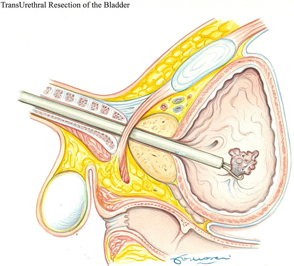
The first approach to bladder cancer is Transurethral
Resection of the Bladder (TURB). The procedure is usually
performed in loco-regional (epidural) anaesthesia. With a
fibre optic instrument introduced trough the urethra, with a
small wire loop all the visible tumor is resected under
vision and extracted
Often it is necessary to maintain an indwelling catheter for
1-5 days, according to the depth of resection, to the number
and extension of lesions.
The procedure obtains an effective local staging of disease.
Prognosis and treatment of bladder cancer differ considerably
according to local stage, whether the tumor is superficial
(not extending beyond mucosa or submucosa) or
muscle-infiltrating (invading the muscular layer of bladder
wall or beyond)

Staging of bladder cancer
TX:Primary tumor cannot be assessed
T0: No evidence of primary tumor
- Ta:Noninvasive papillary carcinoma
- Tis:Carcinoma in situ: "flat tumor"
T1: Tumor invades subepithelial connective tissue T2: Tumor invades muscle
- pT2a: Tumor invades superficial muscle (inner half)
- pT2b: Tumor invades deep muscle (outer half)
T3: Tumor invades perivesical tissue
- pT3a: Microscopically
- pT3b: Macroscopically (extravesical mass)
T4: Tumor invades any of the following: prostate, uterus, vagina, pelvic wall, or abdominal wall
- T4a: Tumor invades the prostate, uterus, vagina
- T4b: Tumor invades the pelvic wall, abdominal wall
Regional lymph nodes (N)
NX: Regional lymph nodes cannot be assessed
N0: No regional lymph node metastasis
N1: Metastasis in a single lymph node, 2 cm or less in
greatest dimension
N2: Metastasis in a single lymph node, more than 2 cm but not
more than 5 cm in greatest dimension; or multiple lymph
nodes, none more than 5 cm in greatest dimension
N3: Metastasis in a lymph node, more than 5 cm in greatest
dimension
Distant metastasis (M)
MX: Distant metastasis cannot be assessed
M0: No distant metastasis
M1: Distant metastasis
In superficial bladder cancer (Ta, T1), which represents
75-80% of all bladder cancers at diagnosis, the prognosis is
usually good, since seldom it tends to progress to muscle
invasion (15%) or to distant metastasis. In case of a
superficial bladder cancer T1 at resection, especially if
high grade (G2-3), it is advisable to perform a second
transurethral resection at 2-4 weeks. In fact, even after
TURB of a skilled urologist, at the second resection residual
tumor can be found up to 40% of cases. Moreover, even if
there is no residual disease, the second resection seems to
improve anyhow the prognosis. In superficial bladder cancer
TURB must be considered a staging and therapeutic procedure.
Local recurrence rate can be high in superficial bladder
cancer, up to 50%-70%. This is due to the fact that bladder
cancer is a multifocal disease, and the tumor may recur in a
bladder site different from the site of resection.
That´s why endovesical therapy with instillations is
often used to reduce recurrence rate. A drug (usually a
chemotherapeutic agent - Mytomicin, Epirubicin, ThioTepa,
Gemcitabine - or an immune system stimulating bacterium -
BCG- Bacillus Calmette Guerin) is introduces by a small
catheter in the bladder once a week, for 6-8 weeks. Local
chemotherapy has not the side effect of a systemic
chemotherapy, since the drug is poorly absorbed through the
bladder wall. Local chemotherapy is effective only at bladder
level, with reduction of recurrence rate. However,
intravesical chemotherapy doesn´t decrease the risk of
progression to muscle-invasive disease. Clinical studies
suggest that in most cases the first cycle of intravesical
chemotherapy could be substituted by an equivalent immediate
single intravesical instillation within 24 hours (even better
within 2-3 hours) from TURB. All intravesical
chemotherapeutic agents have nearly similar efficacy.
However, promising results have been obtained with the recent
introduction of Gemcitabine.
In aggressive superficial bladder cancer (high grade or with
multiple recurrences), the best treatment option is local
immunotherapy with BCG. It is a mild form of the Tubercolosis
Bacterium, used for vaccination. Today it is the most
effective intravesical agent known. It is especially valuable
in high risk superficial cancer (G3, Carcinoma in Situ).
Possible side effects are usually stronger with BCG therapy,
and they may include fever, bladder irritative symptoms,
haematuria, and very rarely urinary or extensive
tuberculosis.
With any kind of intravesical treatment (BCG or chemotherapy)
the literature has shown that the efficacy is improved by
maintenance protocols (repeated cycles after the first ones)
at scheduled intervals.
Though superficial, high grade superficial bladder cancer
(G3T1) deserves special care. This tumor when treated with
transurethral resection only, has a 50% progression risk. BCG
therapy reduces the progression risk to 10-20%, according to
different series.
After TURB f a superficial bladder cancer, patients should
undergo regularly cystoscopy, according to the following
schedule: every 3 months in the first 2 years, every 6
months from the third to the fifth year, and once a year from
then on. To date, no test has shown to be as effective as
cystoscopy to detect cancer recurrence. Bladder cancer
infiltrating the muscular wall of the bladder requires an
open procedure, with removal of the bladder and prostate in
the male and of uterus in the female patient (radical
cystectomy).
All the regional (pelvic) lymph nodes must be dissected and
removed. An extended lymph node dissection (minimum 20 nodes)
seems to improve survival. The operation involves a urinary
diversion, which means a way to expel the urine outside the
body once the bladder has been removed. In majority of case
it is possible to create a new bladder with bowel tissue. A
30-60 cm segment of bowel is isolated, opened, and
reconfigured in spheroid shape. The ureters are sutured to
the new bladder; an anastomosis is created between the
bladder and the residual urethral stump.
After the procedure, the patient can void through the
urethra, with normal self-perception.
However, nighttime´s urinary leakage can occur in
25-30% of cases. A stricture (narrowing) of the suture
between the ureter and the neobladder occurs in 5-15% of
patients, and may require another surgical procedure to avoid
renal damage.
In women there is also the risk of a hyperdistension of the
new bladder, with increase of storage capacity and the risk
of chronic urinary retention.
In poor conditions patients, in patients with impaired renal
function, or with disease extending to the bladder neck
and/or the prostatic urethra, a neobladder is
contraindicated. In these cases an incontinent urinary
diversion, the ileal conduit, is performed. A small
pre-terminal ileal loop is isolated, and the ureters are
sutured to one end. The other end of the loop is sutured
directly to the skin. With this procedure there is no
continence, and the urine is constantly drained into a stoma
bag applied on the skin through an adhesive plate. Though
aesthetically less attractive, this diversion has a lower
risk of complications.
To avoid the discomfort of an incontinent diversion, and when
it is not possible to use the urethral sphincter for the
continence, alternative procedures can be used to obtain
continent reservoirs, like the ileocecal valve,
intussusception of the ileal loop, or reconfiguration of the
ileal loop (Montie procedure) or of the appendix. However,
these diversions require self- catheterization, and the
mechanisms of artificial continence are not always reliable
and effective.
In the last 3-4 years, in laparoscopically trained center,
laparoscopic radical cystectomy is expanding. As laparoscopic
radical prostatectomy, the operation is performed through a
small hole in the abdominal wall, where a fibre optic is
inserted. The abdomen is distended with CO2. Through
additional holes (ports) further instruments are introduced,
and the procedure is guided by a microcamera. Once the
bladder has been isolated, through a small 7-8 cm incision
the bladder is removed and the urinary diversion is performed
in open fashion. Laparoscopic radical cystectomy
can´t be considered a standard procedure yet, but it
can be done in specialized centers.
Five years survival after radical cystectomy for muscle
infiltrating bladder cancer is 50% in older series, but can
reach 80% in recent reports.
In patients at high surgical risk, or refusing surgery, or in
highly selected (single lesion, no residual disease at a
second transurethral resection) and motivated patients, non
invasive protocols with combined approach (radiotherapy and
chemotherapy) may be considered. With this modality, in
selected patients five-years survival is about 50%, and
nearly 40-60% of these patients can preserve the bladder.
However, it must be kept in mind that such protocols are not
standard, and have to be considered experimental.
In cases wit metastatic disease (bone, liver, lung),
chemotherapeutic schedules may provide a prolonged survival,
but the prognosis is usually poor and just a few patients are
still alive at 3 years.