
Kidney Cancer
Kidney cancer is 3% of all tumors in the adult. In
children Wilms tumor is 6% of all pediatric tumors.
Kidney cancer incidence is higher in western countries.
Cigarette smoking is a recognized risk factor. Other factors
include excess of phenacetin-containing drugs, and dialysis.
Family history and a rare congenital syndrome (von
Hippel-Lindau) are related to an increased cancer frequency.
The incidence peak is between 50 and 70 years, with a
male:female ratio of 2:1.
Renal cell carcinoma accounts for about 85% of all renal
cancers. Less usual, but not uncommon, urothelial cancer may
affect the urinary mucosa of the renal excretory way

In 50% of cases a bladder cancer can be associated. Often the first symptom of renal cancer is haematuria (blood in urine). Less specific symptoms include flank weight, fever, and hypertension. In advanced cancer, fatigue and lower limbs edema may occur. Sudden onset of a significant varicocele (dilatation of the vein of the testis) in adult should always be investigated, for the possible vein compression due to renal cancer. In the era of non invasive diagnostic tools, is not rare the incidental finding of a renal cancer with abdominal ultrasound or CT scan.

When a renal cancer is suspected, usually the first approach is a ultrasound of the kidney. However, the main tests for proper diagnosis and clinical staging of renal cancer are abdominal CT scan

or Magnetic Resonance; cancer site and extension can be
evaluated with precision. Since renal cancer can spread to
lung and brain, CT scan should be extended to chest and
brain.
Bone metastases arise in the late course of disease, but if
they are suspected a bone scan must be performed.
Rarely kidney cancer can appear with a cystic aspect.
Because benign renal cysts are very common, sometimes the
diagnosis is not easy. A cyst with thick wall, with solid
tissue inside the lumen, with calcifications, should raise
the suspect of a cancer, especially if they grow fast. In
these cases, they deserve surgical exploration.
TNM classification of kidney cancer
Prumary Tumor (T)
|
TX |
Primary tumor cannot be assessed (information not available). |
|
T0 |
No evidence of primary tumor. |
|
T1a |
Tumor is 4 cm (about 11/2 inches) or less in diameter or and is limited to the kidney. |
|
T1b |
Tumor is larger than 4 cm but not larger than 7 cm (about 2¾ inches) in diameter and is limited to the kidney. |
|
T2 |
Tumor is larger than 7 cm in diameter but is still limited to the kidney. |
|
T3a |
Tumor spread into the adrenal gland or into fatty tissue around the kidney, but not beyond Gerota's fascia. |
|
T3b |
Tumor spread into renal vein and/or the part of the vena cava, still within the abdomen. |
|
T3c |
Tumor spread into vena within the chest or invading the wall of the vena cava. |
|
T4 |
Tumor spread beyond Gerota's fascia |
Regional lymph nodes (N)
|
NX |
Regional lymph nodes cannot be assessed (information not available). |
|
N0 |
No spread to regional lymph nodes. |
|
N1 |
Spread to 1 regional lymph node. |
|
N2 |
Spread to more than 1 regional lymph node. |
Distant metastasis (M)
|
MX |
Presence of distant metastasis cannot be assessed (information not available). |
|
M0 |
No spread to distant lymph nodes or other organs. |
|
M1 |
Distant metastasis is present; includes spread to distant lymph nodes and/or to other organs (such as the lungs, bones, or brain). |
Stage
|
Stage I |
|
|
T1a-T1b, N0, M0: The tumor is 7 cm or less in diameter and limited to the kidney. There is no spread to lymph nodes or distant organs. |
|
|
Stage II |
|
|
T2, N0, M0: The tumor is larger than 7 cm in diameter but is still limited to the kidney. There is no spread to lymph nodes or distant organs |
|
|
Stage III: Different combinations of T and N categories are included in this stage. |
|
|
T3a-T3c, N0, M0: The main tumor has reached the adrenal gland, the fatty tissue around the kidney, the renal vein, and/or vena cava It has not spread beyond Gerota's fascia. There is no spread to lymph nodes or distant organs. |
|
|
T1a-T3c, N1, M0: The main tumor has any size and may be outside the kidney, but it has not spread beyond Gerota's fascia. The cancer has spread to 1 regional lymph node but has not spread to distant lymph nodes or other organs. |
|
|
Stage IV: Several combinations of T, N, and M categories are included in this stage. |
|
|
T4, N0-N1, M0: The main tumor has invaded beyond Gerota's fascia. It has spread to no more than 1 regional lymph node. It has not spread to distant lymph nodes or other organs. |
|
|
Any T, N2, M0: The main tumor has any size and may be outside the kidney. The cancer has spread to more than 1 regional lymph node but has not spread to distant lymph nodes or other organs. |
|
|
Any T, Any N, M1: The main tumor can be any size and may be outside the kidney. It may or may not have spread to regional lymph nodes. It has spread to distant lymph nodes and/or other organs. |
Radical nephrectomy (removal of the kidney
with surrounding fat tissue) is the standard treatment of
renal cancer. Nephrectomy can be performed either with open
or laparoscopic approach.With laparoscopy, CO2 is
insufflated in the abdomen. A microcamera and a few ports
(trocars), 5 to 12 mm in diameter, are introduced through
small holes in the abdominal wall
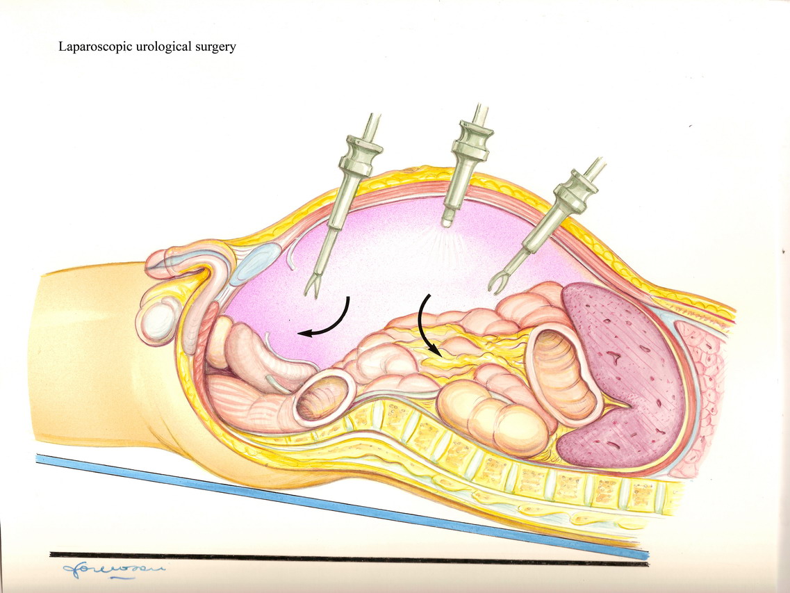
The operation is performed with delicate
and long instruments introduced through the trocar, guided on
a screen.
Since 2006 the guidelines European Association of Urology
advocate laparoscopic nephrectomy for the treatment of
localized kidney cancer, for decreased surgical trauma and
faster recovery.
Ten-year oncological results of laparoscopy radical
nephrectomy are comparable to the ones of open surgery. Even
when the tumor extends to the vena cava, if the vein wall is
not infiltrated and the tumor is completely removed, healing
chance can be high.
Usually, if the controlateral kidney is healthy, the quality
of life after nephrectomy is excellent.
However, complete removal of the kidney for cancer is not
always necessary. For peripheral tumors, not larger than 4 cm
(even if sometimes tumors up to 7 cm can be treated), and not
involving the vascular hilum of the kidney, a partial
nephrectomy is a feasible option. The tumor is resected with
a thin layer (between 3 and 5 mm) of healthy tissue all
around. The renal vessels are often temporary occluded, to
reduce blood loss during tumor resection. The kidney tissue
is then sutured. This technique is named partial
nephrectomy.
In long term studies, partial nephrectomy has shown to be
equivalent to radical nephrectomy at 15 years of followup.
This procedure too can be performed with laparoscopic
approach, but the technical complexity requires laparoscopic
skilled hospitals.
Recently, cryotherapy or radiofrequency treatment has been
proposed for small peripheral tumors in very old or high risk
patients. Small probes must be inserted in the
tumor, trough a percutaneous or a laparoscopic
approach. The tumor is thhn
"freezed" at very low temperature, or heated with
electromagnetic waves.
Though promising, these procedures should be considered
investigative. After radical nephrectomy for non-metastatic
kidney cancer, 5-years survival is 85%.
Nearly 20-30% of patients after radical nephrectomy will
develop local or distant disease recurrence.
About 25% of patients with kidney cancer present distant
metastasis at diagnosis. Some reports have shown that for
metastatic disease, nephrectomy plus immunotherapy can be a
choice for patients with good performance status and
asymptomatic. However, even if survival may be prolonged, the
prognosis in these patients is often poor.
In selected cases of single, amenable of surgical removal
metastasis, nephrectomy plus metastasis resection is a
feasible option.