
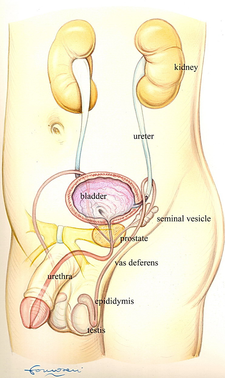
Prostate Cancer
Prostatic cancer is the first tumour for incidence (29% of
all cancers) and the second cause of cancer death (13% of all
cancers, second to lung cancer only) for men in western
countries.
However, the natural history of the tumour may be slow.
Nearly one man out of 6 will be diagnosed with prostate
cancer, but the disease mortality is 3%. So it is estimated
that only 20% of men with a prostate cancer
diagnosis will die of the tumour, while the
remaining 80% will die with the tumor, but for
another reason.At the age of 80 about 20-40% of men
have some foci of prostate cancer. The disease risk is
increased by a diet rich with fat.Recently it has been shown
that a virus might promote the onset of prostate cancer.
The course of the disease is depending upon the grade of the
tumour. Prostate cancer grading is based on a score
(Gleason), going from 2 (well differentiated tumor) to 10
(undifferentiated tumor). In recent reports, it has been
established that for clinical purpose the histology grade
should be limited to Gleason 5 (low grade), Gleason 6
(low-intermediate grade), Gleason 7 (intermediate grade)
Gleason 8-10 (high-very high grade). Fifteen year mortality
for untreated cancer is 9% for low grade, 28% for
intermediate grade, and 58% for high grade disease.
In younger patients, life expectancy for untreated cancer is
decreased by 4-5 years and 6-8 years for intermediate and
high grade, respectively. In patients younger than 65 years
with localized cancer (no spread to lymph nodes and no
distant metastasis), mortality for untreated cancer can be as
high as 65%.In my opinion, patients with prostate cancer
younger than 70 years should undergo definitive treatment;
patients between 70 and 75 years may receive definitive
treatment according to grade and stage of disease, and to
general health status; palliative treatments should be
reserved for patients older than 75 years. Surgery should be
offered to all patients with intermediate or high grade
localized cancer and a life expectancy at least of 10-15
years.
Diagnosis
PSA
Prostate cancer is asymptomatic for a long time. When
symptoms like urinary obstruction or bone pain arise, the
disease is too advanced and beyond the chance of definitive
therapy.
The diagnosis of prostate cancer has changed dramatically in
the last fifteen years, due to the introduction of the most
useful biological marker in oncology: the Prostate Specific
Antigen (PSA). The PSA is a protein that, for practical
clinical use, can be considered of exclusive prostatic
origin. Usually the concentration is very high in prostatic
secretions and in the semen , and extremely low in the
circulating blood.. PSA is produced by normal, hyperplasic,
and cancer prostatic cells (also from distant metastasis).
Any change of prostatic cellular structure can induce an
elevated PSA, due to increased "leakage" of PSA from the
prostatic cells to the blood vessels. Acute, and sometimes
chronic infection of the prostate may increase the PSA, maybe
for increased vessel permeability during inflammation. An
indwelling catheter, transurethral procedures, and prostatic
massage can all cause a transient elevation of PSA. Prostatic
biopsy obviously produces a temporary rise of PSA. PSA should
be measured again only 4-6 weeks after prostatic biopsy.
The half-life of PSA in the blood is about 3 days. Benign
prostate enlargement can cause a moderate PSA increase,
especially in very large prostates.
The main clinical interest of PSA is in the diagnosis,
staging, prognosis and treatment evaluation of prostate
cancer.
For early prostate cancer diagnosis, every male at the age
of fifty should start PSA measurement on annual basis. The
screening should start at the age of forty for afro-americans
and for patients with a family history of prostate cancer.
The normal reference range of PSA is between 0.1 and 4.0
ng/ml. When PSA is beyond 4.0 ng/ml, the chance of having a
prostate cancer is 33-51%, according to different series
and/or the biopsy method. In the PSA range between 4.0 and
10.0, the incidence of cancer is 24-47%.
Some Authors have proposed to decrease the cut-offto 2.5
ng/ml. When PSA is between 2.5 and 4.0%, the incidence of
cancer is 24-27%. Though an increased number of negative
biopsies would be performed with this limit, a higher number
of cancers would be detected, particularly in young patients.
Of course these data are not absolute. For instance, a PSA
value of 3.9 ng/ml, though in the normal range, should anyhow
be investigated.
For diagnostic purpose, it is very important the way the PSA
changes along time (PSA velocity). An increase of 1ng/ml/year
is considered suspicious. A PSA that in 2 years interval
moves from 2.0 to 6.0, is probably more wary of a PSA which
has been stable around 9 ng/ml in the last 5 years, in a
prostate with a very large adenoma.
The free PSA is a portion of the total PSA very sensitive
for prostate cancer diagnosis. A ratio of free and total PSA
below 18% is suggestive of prostate cancer, in the range of
total PSA between 2.5 and 10.0 ng/ml.
New molecular forms of PSA are under investigation, with
promising results.
Digital rectal examination (DRE)
Digital rectal examination (DRE) is a simple, fast and cheap
test for prostate cancer diagnosis, thou less sensitive than
PSA. With DRE only, up to 45%-70% of prostate cancers can be
missed. However, since some tumors may present with normal
PSA, DRE should be performed in every man after 50.
Transrectal ultrasound (TRUS)
A good imaging of the prostate can be obtained through
transrectal ultrasound (TRUS). Usually cancer can be seen as
a hypoechoic (darker than the surrounding tissue) nodule or
peripheral area.

|

|
Nevertheless, up to 40% of cancers are not seen by
transrectal ultrasound. The tumor extension may be
underestimated by TRUS. When the disease is extracapsular (it
has reached and penetrated the prostate capsule), TRUS is not
able to predict it in 40-50% of cases. However, when an
extracapsular disease is seen by ultrasound, the tumor has a
80-90% chance of extendingbeyond the prostate capsule. Though
it has some limits, transrectal ultrasound can be helpful for
the detection and staging of prostate cancer.
Magnetic Resonance (MR) with Endorectal coil
This recent and promising technique for detecting and
locally staging prostate cancer is expanding and becoming
very reliable. In the next future it could become a key point
for decision making in the treatment of prostate cancer.
I personally prescribe the test to all men candidate to
radical prostatectomy.
With endorectal MR there is a high definition imaging of the
prostate and surrounding tissues.
The latest instruments can provide a metabolic analysis of
the prostatic tissue, with special attention to Choline and
Citrate concentrations, that can be altered in tumors.
Prostate biopsy
Prostate biopsy is the only way to prove the presence of a
prostate cancer. The biopsy is performed with a specially
designed cutting needle; with a double advancement a tiny
sample of prostatic tissue is obtained. The biopsy is
performed through a transrectal (the needle is passed through
the rectal wall) or a transperineal (through the skin between
the scrotum and the anus) approach.
Digital guided biopsy is outdated and the procedure is
performed today under transrectal ultrasound control. In most
western countries the transrectal approach is preferred.
Since a large percentage of tumors are not clearly visible by
ultrasound, the samples (cores) are obtained according to
random schemes. Of course all hypoechoic lesions are included
in the biopsy.
Some authors recently have suggested evaluation of prostatic
tissue through Doppler transrectal ultrasound, to identify
areas with increased blood supply, suspicious for cancer.
Further studies are needed to confirm this approach.
Sextant (with 6 cores) transrectal biopsy has been widely
used in the nineties. However, in the last decade several
studies have questioned the sextant technique, and new
protocols with increased number of cores have been
introduced. Six cores are not considered enough for adequate
prostate cancer detection, especially in large prostates.
Nowadays a good prostate biopsy should include at least 8
cores, with optimal number of cores between 10 and 12. In
patients with increasing PSA and one or more negative
biopsies, some urologist propose a saturation biopsy, with
protocols that can obtained sometimesmore than 30 cores.
Personally since 1997 I´ve useda "fan" technique, with
12 cores obtained through a perineal approach. The results in
more than 2000 patients are satisfying. Prostate cancer has
been detected in 47% and 45% of cases with PSA above 4.0 and
PSA between 4.0 and 10.0 ng/ml, respectively.
Since in my series nearly half the patients with PSA above
4.0 ng/ml have a prostate cancer, I recommend a 10-12 core
biopsy in all patients with increased PSA.
Prostate biopsy is an office procedure. With transrectal
approach the local anesthesia is not strictly required, but
several studies have shown that with local anesthesia, the
procedure is better accepted.
The local anesthesia is needed with transperineal approach.
Temporary haematuria (blood in the urine) for 7-10 days and
haemospermia (blood in the semen) for 2-3 months are not
uncommon after prostate biopsy.
PrimaryTumor (T)
TX Primary tumor cannot be assessed
T0 No evidence of primary tumor
T1 Clinically inapparent tumor not palpable
nor visible by imaging
- T1a Tumor incidental histologic finding in 5% or less of tissue resected (in prostatectomy)
- T1bTumor incidental histologic finding in more than 5% of tissue resected
- T1cTumor identified by needle biopsy (e.g., because of elevated PSA)
T2 Tumor confined within prostate
- T2a Tumor involves one-half of 1 lobe or less
- T2bTumor involves more than one-half of 1 lobe but not both lobes
- T2cTumor involves both lobes
T3 Tumor extends through the prostate capsule
- T3a Extracapsular extension (unilateral or bilateral)
- T3b Tumor invades seminal vesicle(s)
T4 Tumor is fixed or invades adjacent
structures other than seminal vesicles: bladder neck,
external sphincter, rectum, levator muscles,and/or pelvic
wall.
Regional lymph nodes (N)
NX Regional lymph nodes were not assessed
N0 No regional lymph node metastasis (lymph
nodes confined to the true pelvis)
N1 Metastasis in regional lymph node(s)
Distant metastasis (M)
MX Distant metastasis cannot be assessed
(not evaluated by any modality)
M0 No distant metastasis
M1 Distant metastasis
- M1a Nonregional lymph node(s)
- M1b Bone(s)
- M1c Other site(s) with or without bone disease
Prostate cancer: Treatment.
After a prostate cancer diagnosis, accurate staging of the
disease is essential. Local staging is achieved trough
Digital Rectal Examination, Magnetic Resonance with Endocoil.
Important staging factors are the value of PSA, the Gleason
score, the percentage of positive cores in the biopsy, and
the fraction of total cancers in all cores. The combination
of these data can provide a reliable staging method. When PSA
is below 10.0 ng/ml and Gleason score is below 7, the chances
of having disease metastases to lymph nodes or to the bone is
very low. All the other patients should undergo a CT or MR of
the pelvis and a bone scan.
Occasionally some tumors are very small (less than 0.5 cc of
volume) and with a Gleason score lower than 6. These tumors
are considered not clinically significant, and can deserve
watchful waiting, rather than aggressive treatment.
Scandinavian studies have proposed a watchful waiting for
moderately aggressive tumors (Gleason score less than 7, PSA
lower than 10.0 ng/ml). Though this approach can be
reasonable in elder patients (70-75 years of age or older),
recent data show that when patients with moderately
aggressive disease are followed for 15-20 years, cancer
mortality is higher than 50%.
Therefore we must be very cautious before proposing watchful
waiting in patients of 65 years or younger.
When the disease is still inside the gland, it can be fully
cured through surgical removal of the gland (radical
prostatectomy). Since prostate cancer is usually slowly
progressing, usually radical prostatectomy should be reserved
to patients with at least 10 years of life expectancy
(generally they are patients 70 years old or younger). In low
grade tumors (Gleason score 5 or less) a conservative
approach can be reasonable, since the chance of tumor
progression at 15 years is rather low. Aggressive tumors
(Gleason 8 or higher) may require radical prostatectomy in
selected patients older than 70, as poorly differentiated
tumors can grow rapidly and progress to metastatic disease in
most patients in 5-6 years.
Open radical prostatectomy can be performed through a
retropubic or a perineal approach. Preoperative hormone
therapy has no influence on radical prostatectomy results,
and today it is considered useless.
Radical prostatectomy is a well standardized procedure. The
average blood loss is between 200 and 1000 cc. When the tumor
doesn´t reach the prostate capsule, the erection nerves
that run on the postero-lateral aspect of the gland, can be
spared. However, even after a nerve-sparing procedure, some
grade of erectile dysfunction can be present in 20-70% of
cases.
Temporary stress urinary incontinence in not uncommon after
the procedure., lasting form a few weeks to several months.
After 6-12 months only 3-5% of patients have still some
degree of incontinence.
Recently a new approach to radical prostectomy, laparoscopy,
is expanding in western countries. The operation is performed
through some small holes in the abdominal wall, with long and
small instruments that are guided by a microcamera
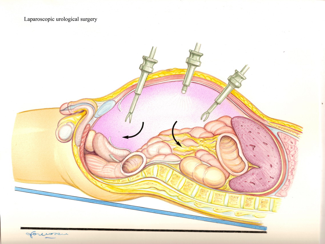
The peritoneal cavity, or the pelvic space anterior to the
prostate, are inflated with C02. The tissues are distended by
the gas pressure. The whole procedure is done with a great
magnification on a screen. The advantages are better
anatomical details and control, the clear visualization of
areas that can´t be seen with the open approach, a
mini-invasive procedure, a lower blood loss (average 100-200
cc). The operation can be performed with conventional
laparoscopy, or with robot assisted laparoscopy. The robot is
a complex apparatus, with robotic arms, guided by a computer
that reproduces the movements of the operator´s hands
outside the body. The surgeon is sitting at a console, where
he/she can have a three-dimensional view of the operating
field. Small and versatile instruments perform the procedure
inside the body. The use of the robot is especially spread in
US, where 75% of prostatectomies are performed by robot
assisted laparoscopy.
The recovery to normal activities is usually faster after
laparoscopic radical prostatectomy. The functional results
(about continence and potency) are at least equal to the ones
of open surgery. The first oncological results (no evidence
of disease) at five years are comparable to the ones obtained
by excellence centers with open radical prostatectomy.
Disadvantages include a long learning curve, especially for
laparoscopy beginners, and a moderately longer operative
time. The learning curve is shorter with the use of the
robot.
If we include only studies with more than 100 cases, more
than 10,000 laparoscopic radical prostatectomies have been
reported in literature. Probably the number of procedures
performed all over the world is 3-4 times higher. The results
are very encouraging and this mini-invasive procedure could
become in the next future the standard of prostatic cancer
surgery.
Today, the surgeon should offer to the patient the technique
he feels confident in, according to his experience and
skills.
When after radical prostatectomy the surgical margins of the
prostate are positive for disease (the cancer reaches the
external limit of the surgical resection), there is a higher
risk of local recurrence of tumor. In this group of patients,
an early radiotherapy (60 Gy) can appreciably improve
prognosis and survival.
The cure rate after radical prostatectomy is depending upon
several factors, including stage at final pathology, Gleason
score, preoperative PSA, preoperative PSA velocity, surgical
margin status.
After radical prostatectomy, patients are followed with PSA
measurement every 3 months.
At 10 years, in nearly 70% of patients no significative PSA
(less or equal to 0.2 ng/ml) can be detected, 20% of patients
have increased PSA without any other clinical sign of
disease, and 10% of patients have local recurrence or
metastatic disease.
A PSA is 0.4 ng/ml or above means recurrence of disease.
When the PSA reaches this value at least 18 months after
surgery, the patient is likely to have a local recurrence,
rather than distant metastasis. When a local recurrence is
present, still 30-45% of patients can be successfully treated
with local radiotherapy.
Radiotherapy
High energy radiation can treat prostate cancer. The
treatment usually last a few weeks, and it is well tolerated.
New techniques allow 3-dimensional imaging of the target (the
gland), with more precise, high dose energy and decreased
side effects on surrounding organs (mainly bladder and
rectum/anus)

Brachitherapy is another radiation treatment modality which
is becoming popular. Brachiterapy is performed releasing some
radioactive seeds in the prostate. The optimal dose is
evaluated through CT scan-based 3-D imaging of the gland.
Ten-year results are promising, apparently equivalent to the
ones of conventional external beam radiotherapy. However the
risk of urethral strictures and rectal fistulas is higher
with brachitherapy compared to external radiotherapy.
Main side effects of external radiotherapy are bladder and
rectum irritative symptoms, at times with diarrhoea,
increased urinary frequency. In only 5% of cases these
symptoms become chronic.
Some degree of erectile dysfunction can occur after
radiotherapy in 40-70% of patients.
Even several years after radiotherapy severe haematuria
(blood in the urine) can occur.
Some authors have proposed to increase radiotherapy efficacy
with concomitant hormone therapy, to be continued for 3
years. The aim of the combined treatment is to decrease the
tumor volume and to increase radiosensitivity of tumor cells.
This approach has proven to be more effective in aggressive
or locally advanced prostate cancers.
Radiotherapy results are not easy comparable to the ones of
radical prostatectomy, since after radiotherapy PSA
doesn´t reach the same levels as after surgery. PSA
reaches the lowest concentration (nadir) usually in 18-24
months. Since prostate cancer is often a slowly
progressing disease, results evaluation needs a followup of
10-15 years. Disease-free survival after radiotherapy for
curable prostate cancer is 75-95% at 5 years and 55-85% at 10
years, according to literature. Anyhow, about 50-60% of
patients after radiotherapy have still cancer at prostate
biopsy; this group is at increased risk of progression and
disease related death.
The results of radiotherapy for locally advanced tumors
(extending beyond capsule, without distant metastases) are
less encouraging, with 15-30% survival at 10 years.
Cryotherapy has been proposed to treat prostate cancer
recurrent after radiotherapy failure. However, a long
followup is needed to assess the results and the risks of the
treatment.
The experience of high frequency radio waves application
(HIFU) for prostate cancer is stll preliminary.
Though radical prostatectomy is feasible after the failure
of radiotherapy, the risk of complications, including urinary
incontinence, is higher
Hormone therapy
Though the effects of androgen deprivation on prostate
cancer are well known, hormonal therapy should be considered
a palliative treatment in very old or with metastatic disease
patients, rather than a definitive therapy.
Oestrogen therapy has been almost abandoned for serious
cardiovascular side effects.
Several drugs are today available (Leuprolide, Goserelin,
Buserelin, Triptorelin) for inhibition of testosterone
secretion (Luteining Hormone Releasing Hormone - LHRH
analogues). They are available in monthly or quarterly
administration. Usually hormone therapy is well tolerated.
Side effects can include erectile dysfunction, mood changes,
loss of muscle tissue, bone demineralization, and hot rushes.
Other drugs used for hormone therapy are antiandrogens
(flutamide, bicalutamide, nilutamide, cyproterone
acetate). They can be used together with LHRH analogues or as
monotherapy. Though side effects on sexual function are less
severe, androgen monotherapy for prostate cancer is not
considered a standard therapy. The association between
an LHRH analogue and an antiadrogen is known as "Total
Androgen Blockade", and it is still debated. Some papers have
reported an increase of median survival of 7 months with
total androgen blockade, but a recent review of the
literature leaves the problem still open. A large study of
EORTC didn´t show any significant advantage when an
antiandrogen was added to the therapy with LHRH analogues.
Intermittent hormone therapy has been proposed. The
rationale is based upon the fact that after an interval of
time some cell clones become resistant to hormone
deprivation. Interrupting the hormone therapy might increase
biological competition between hormone dependent and not
hormone dependant cells, delaying the onset of hormone
refractory disease. Long term randomized trials are required
to asses the efficacy, if any, of this approach.
The association of hormone therapy and radiotherapy seems to
improve survival for locally advanced prostate cancer.
Generally hormone therapy is given to patients with
metastatic disease, or not otherwise treatable, or after the
failure of surgery and/or radiotherapy.
For metastatic disease, early hormone treatment delays
disease progression and prolongs survival, compared to
delayed (at the onset of symptoms) treatment. It is
uncertain if elder patients with localized disease could
benefit from early hormone therapy. Again, in a study from
EORTC, in these patients there is no results´difference
between early and delayed hormone therapy.
How I treat prostate cancer
I consider radical prostatectomy the best therapeutic option
to obtain definitive cure for intracapsular prostate cancer.
In patients younger than 71 years and with Gleason score 6 or
higher, unless there is clinically insignificant cancer,
radical prostatectomy is my first choice. For patients
between 71 and 75 years old, radical prostatectomy can be an
option in motivated patients, in patients with long life
expectancy, or with high grade cancer (Gleason score 7 (4+3),
or 8-10) deserving aggressive therapy, since this last group
could experience disease progression within 5-8 years.
In patients with Gleason score 5 and localized prostate
cancer, radical prostatectomy is a good option for patients
59 years old or younger, while expecting approach can be
reasonable only for patients older than 60 years, due to the
fact that cancer progression is unlikely to occur on a long
time period. These patients, however, must be followed
strictly to assess if the disease is very slow, or if there
is an evolution trend. When the tumor is localized,
radiotherapy is a good option for patients of age
75-80, for patients between 70 and 75 years with
Gleason score 7 (3+4) or less, or for patients with extensive
extracapsular disease, where the chance of surgical
cure is low. Hormone therapy is my treatment for metastatic
disease, or recurrence after radical prostatectomy and/or
radiotherapy. In very old patients it can be an option, to
start immediately for advanced disease. The hormone treatment
can be delayed in very old patients with localized
asymptomatic prostate cancer.